Home » Without Label » Arteries Diagram - Ultrastructure Of Blood Vessels Arteries Veins Teachmeanatomy - Arteries of the head and neck diagram art print vintage anatomy art print on tea stained paper dog art dog s wfh office art.
Arteries Diagram - Ultrastructure Of Blood Vessels Arteries Veins Teachmeanatomy - Arteries of the head and neck diagram art print vintage anatomy art print on tea stained paper dog art dog s wfh office art.
Arteries Diagram - Ultrastructure Of Blood Vessels Arteries Veins Teachmeanatomy - Arteries of the head and neck diagram art print vintage anatomy art print on tea stained paper dog art dog s wfh office art.. The coronary arteries wrap around the outside of the heart. Learn vocabulary, terms, and more with flashcards, games, and other study tools. An artery (plural arteries) (from greek ἀρτηρία (artēríā) 'windpipe, artery') is a blood vessel that takes blood away from the heart to one or more parts of the body (tissues, lungs, brain etc.). The heart muscle also needs it. By definition, an artery is a vessel that conducts blood from the heart to the periphery.
Most arteries carry oxygenated blood; These arteries and their branches supply all parts of the heart muscle with blood. Arteries carry blood away from the heart in two distinct pathways: Only 3 available and it's in 2 people's carts. Arteries, veins, and the heart are the main parts of the system.
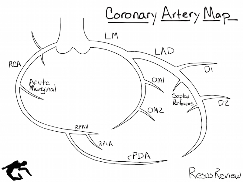
Coronary Artery Diagramming Resus Review from resusreview.com Other arteries of the neck. Smartdraw includes 1000s of professional healthcare and anatomy chart templates that you can modify and make your own. 5 out of 5 stars. The narrowed arteries are at higher risk for complete blockage from a sudden. Over the years, cholesterol plaques can narrow the arteries supplying blood to the heart. Ascending aorta, aortic arch, thoracic aorta, and abdominal aorta. The coronary arteries wrap around the outside of the heart. Arteries are components of the cardiovascular system.
The aorta branches into a network of smaller arteries that extend throughout the body.
This buildup is called plaque. It can also help them in getting an overview of artery vs. Diagram showing the effects of atherosclerosis on an artery. Creating a freehand diagram of arteries and veins can be troublesome. Ascending aorta, aortic arch, thoracic aorta, and abdominal aorta. The defect is associated with narrowing of the trachea (windpipe) and bronchi (airways). Arteries are components of the cardiovascular system. The arteries' smaller branches are called arterioles and capillaries. Finally, the smallest arteries, called arterioles are further branched into small capillaries, where the exchange of all the nutrients, gases and other waste molecules are carried out. Is associated with venipuncture, it is done mainly by phlebotomists, nurses, emts and doctors. It ends at the anterior and posterior tibial arteries. Arteries, veins, and the heart are the main parts of the system. To understand the system, the students need to create their diagrams.
This buildup is called plaque. Finally, the smallest arteries, called arterioles are further branched into small capillaries, where the exchange of all the nutrients, gases and other waste molecules are carried out. The two exceptions are the pulmonary and the umbilical arteries, which carry deoxygenated blood to the organs that oxygenate it (lungs and placenta. There is a printable worksheet available for download here so you can take the quiz with pen and paper. It is located in the middle cavity of the chest, between the lungs.
40 3b Arteries Veins And Capillaries Biology Libretexts from textimgs.s3.amazonaws.com 15 diagram of main arteries. The right and left subclavian arteries give rise to the thyrocervical trunk. After receiving blood directly from the left ventricle of the heart, the. The cardiovascular system consists of the heart, blood vessels, and the approximately 5 liters of blood that the blood vessels transport. The plaque can also burst, leading to a blood clot. In this image, you will find external carotid artery, internal carotid artery, vertebral artery, aorta and arch, pulmonary artery, cardiac artery, thoracic aorta, celiac trunk, superior mesenteric artery, renal artery, gonadal artery, inferior mesenteric artery, common iliac artery, external iliac artery. Arteries, veins, and the heart are the main parts of the system. Arteries of the leg diagram.
By definition, an artery is a vessel that conducts blood from the heart to the periphery.
Create healthcare diagrams like this example called arteries and veins of the arm in minutes with smartdraw. The two exceptions are the pulmonary and the umbilical arteries, which carry deoxygenated blood to the organs that oxygenate it (lungs and placenta. The tunica medica, which is the very muscular middle layer in arteries, is thinner and less muscular in veins. By definition, an artery is a vessel that conducts blood from the heart to the periphery. More artery diagrams are posted in the following 101 diagramss below. Arteries, veins, and the heart are the main parts of the system. John bavosi/science photo library/getty images. Other arteries of the neck. Arteries and arterioles carry oxygenated blood _____ from the heart to the body. There is a printable worksheet available for download here so you can take the quiz with pen and paper. Veins are the blood vessels present throughout the body. From this trunk, several vessels arise, which go on to supply the neck. It can also help them in getting an overview of artery vs.
There is a printable worksheet available for download here so you can take the quiz with pen and paper. Arteries of the lower limb thigh leg foot the main artery of the lower limb is femoral artery it is a continuation of the external iliac artery terminal branch of the abdominal aorta the arteries and veins of the leg smartdraw arteries and veins of the leg create healthcare diagrams like this example called arteries and veins of the leg in minutes with smartdraw. Most arteries carry oxygenated blood; In this image, you will find right gastric artery, common hepatic artery, celiac trunk, left gastric artery, splenic artery, splenic vein, pancreas, suprarenal vein, renal vein, renal artery, inferior mesenteric vein , gonadal vein, gonadal artery, two alternative position of artery, left colic artery. The arteries' smaller branches are called arterioles and capillaries.
2 Carotid Ultrasound Anatomy 123 Sonography from www.123sonography.com Arteries carry blood away from the heart in two distinct pathways: From this trunk, several vessels arise, which go on to supply the neck. Cholesterol can build up in the arteries as a person gets older, and this is more likely for people who have diets. The arteries' smaller branches are called arterioles and capillaries. Other arteries of the neck. After receiving blood directly from the left ventricle of the heart, the. The two exceptions are the pulmonary and the umbilical arteries, which carry deoxygenated blood to the organs that oxygenate it (lungs and placenta. Most arteries carry oxygenated blood;
More artery diagrams are posted in the following 101 diagramss below.
Only 3 available and it's in 2 people's carts. Arteries of the lower limb thigh leg foot the main artery of the lower limb is femoral artery it is a continuation of the external iliac artery terminal branch of the abdominal aorta the arteries and veins of the leg smartdraw arteries and veins of the leg create healthcare diagrams like this example called arteries and veins of the leg in minutes with smartdraw. Coronary arteries supply blood to the heart muscle. The two exceptions are the pulmonary and the umbilical arteries, which carry deoxygenated blood to the organs that oxygenate it (lungs and placenta. Finally, the smallest arteries, called arterioles are further branched into small capillaries, where the exchange of all the nutrients, gases and other waste molecules are carried out. Smartdraw includes 1000s of professional healthcare and anatomy chart templates that you can modify and make your own. The coronary arteries wrap around the outside of the heart. This buildup is called plaque. In this image, you will find right gastric artery, common hepatic artery, celiac trunk, left gastric artery, splenic artery, splenic vein, pancreas, suprarenal vein, renal vein, renal artery, inferior mesenteric vein , gonadal vein, gonadal artery, two alternative position of artery, left colic artery. Creating a freehand diagram of arteries and veins can be troublesome. It can also help them in getting an overview of artery vs. Arteries carry blood away from the heart in two distinct pathways: The arteries' smaller branches are called arterioles and capillaries.